Having an External Ventricular Drain (EVD)
An external ventricular drain (EVD) is a thin tube that drains extra cerebrospinal fluid (CSF) from the brain. The extra fluid may be caused by illness or injury. Too much fluid raises the pressure in and around the brain. An EVD helps reduce pressure in the brain to a safe level.
How to say it
ehks-TER-nuhl vehn-TRIH-kyoo-ler
Why is an EVD needed?
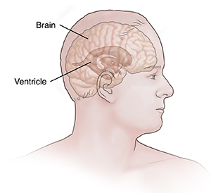
CSF is a liquid that is in the brain cavities (ventricles) and around the brain and spinal cord. CSF is made by cells in the brain’s ventricles. The ventricles are spaces in the middle of the brain. Too much CSF can build up in the ventricles if the flow of fluid is blocked by a birth defect or an illness such as meningitis or bleeding. This is called hydrocephalus. In some cases, the ventricles may fill with blood because of an injury, a stroke, or another cause. Hydrocephalus increases pressure in the brain. Too much pressure in the brain can cause headaches, nausea, vomiting, confusion, loss of consciousness, and brain damage. An EVD drains some of the CSF or blood to restore a safe level of pressure in the brain. It also allows the healthcare team to record the brain pressure (intracranial pressure).
How does an EVD work?
A healthcare provider puts a thin, flexible tube (catheter) through the top of the skull into the center of the brain during a surgery or at bedside. Some EVDs are covered with an antibiotic. The tube is attached to a pouch nearby. The tube drains the extra fluid into the pouch. The amount of fluid is measured as it drains. The EVD is left in place as long as needed. This may be several days, a week, or longer.
Risks of an EVD
All procedures have risks. The risks of an EVD include:
-
Problems placing the tube in the right place in the brain
-
Infection of CSF with bacteria or fungi
-
Too much bleeding where the tube is inserted. This can happen when it is placed or when it is removed.
-
Brain damage where the EVD is placed
-
Blood clot that blocks the tube
-
Too much fluid drained away
-
EVD doesn't work as it should
During EVD placement
A neurosurgeon, a neurosurgery resident, or an intensivist will do the procedure. The surgery can be done in several ways. Ask the healthcare provider about the details of this surgery. In general, you can expect the following for your loved one:
-
They will be given medicine to help them relax (sedation), or medicine that lets them sleep through the surgery (general anesthesia). Or the medical team may inject anesthesia into the scalp. Antibiotics may be given during and after the surgery. This is to help prevent infection.
-
The healthcare team will trim the hair on top of the person's head. The surgeon makes a small cut (incision) in the skin.
-
Using a special drill, a surgeon drills a small hole in the bone of the skull. They then make a small incision in the layers covering the brain.
-
The surgeon puts a thin tube into the incision and then through the brain. In some cases, the surgeon may use computer-assisted imaging to help the tube reach the right place in the brain.
-
The surgeon then uses a small stitch (suture) on the skin of the scalp to hold the tube in place. A sterile dressing is put over the incision and the stitch.
-
The surgeon connects the tube to the pouch.
While your loved one has an EVD
The healthcare team will check the amount of fluid in the pouch often. While an EVD is in place, antibiotics may be given to help prevent infection. The CSF may also be checked regularly for signs of infection. Tell the healthcare team if your loved one has any symptoms that come back or get worse, such as nausea or confusion.
When is an EVD removed?
The EVD is removed as soon as the healthcare team decides it’s safe to take out. In some cases, an EVD is replaced with a different tube called a VP (ventriculoperitoneal) shunt. This is an internal tube that drains the extra fluid to a place inside the belly (abdomen). This kind of tube can stay in place for a longer time. A VP shunt is put in place during a separate procedure. The healthcare team will tell you more if your loved one needs this.
Online Medical Reviewer:
Anne Fetterman RN BSN
Online Medical Reviewer:
Luc Jasmin MD
Online Medical Reviewer:
Raymond Kent Turley BSN MSN RN
Date Last Reviewed:
12/1/2022
© 2000-2024 The StayWell Company, LLC. All rights reserved. This information is not intended as a substitute for professional medical care. Always follow your healthcare professional's instructions.